Ragon Institute Imaging Core
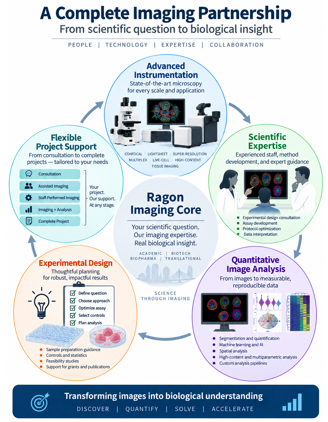
The Ragon Institute Imaging Core provides investigators with access to state-of-the-art microscopy technologies, expert consultation, and advanced image analysis to accelerate discoveries in immunology, infectious diseases, cancer, neuroscience, and cell biology. As an MGH Core Facility, the Core serves both the Ragon Institute and the broader MGH research community by supporting imaging-based research from experimental design through data acquisition, quantitative analysis, and interpretation.
Located on the 4th floor of 600 Main Street (Rooms 434 and 436, Cambridge, MA), the Imaging Core offers a comprehensive portfolio of advanced imaging platforms spanning widefield, confocal, spinning-disk confocal, super-resolution, light-sheet, multiplex tissue imaging, and live-cell microscopy. Our staff provides consultation on experimental design, fluorophore selection, multiplex imaging strategies, image analysis, and quantitative microscopy, while also offering instrument training and technical support.
Imaging Platforms
The Imaging Core provides access to a diverse collection of imaging systems designed to accommodate applications ranging from routine fluorescence imaging to advanced multidimensional microscopy.
Leica STELLARIS Laser Scanning Confocal Microscopes provide high-sensitivity spectral confocal imaging using White Light Laser excitation and tunable spectral detection. These systems enable flexible fluorophore selection, spectral imaging, linear unmixing, and highly multiplexed fluorescence imaging while minimizing spectral overlap and autofluorescence.
Nikon Inverted Spinning-Disk Confocal Microscope with CrestOptics CICERO Confocal Head delivers high-speed, low-phototoxicity imaging for live-cell experiments, long-term time-lapse studies, intracellular dynamics, and high-throughput imaging applications. The system is ideally suited for rapid volumetric imaging and dynamic biological processes.
TissueFAXS SLQ Confocal Slide Scanner is a high-throughput spinning-disk confocal whole-slide imaging system capable of automated acquisition of up to 120 slides. It supports brightfield, fluorescence, and confocal imaging with z-stacks, making it ideal for large tissue studies and quantitative cellular analysis.
TissueFAXS Plus Whole-Slide Scanner provides rapid brightfield and fluorescence scanning of up to 8 slides sequentially, supporting multiplex imaging of up to 10 fluorescent markers and iterative staining workflows for highly multiplexed tissue analysis. The system incorporates a Lumencor Spectra light engine and supports quantitative image analysis using TissueQuest, HistoQuest, and StrataQuest software.
ZEISS Celldiscoverer 7 with LSM 900 and Airyscan 2 combines automated widefield imaging with laser scanning confocal microscopy and Airyscan super-resolution. The platform supports imaging of slides, multiwell plates (up to 1536 wells), chamber slides, and culture dishes, with capabilities including 2D, 3D, and 4D imaging, long-term live-cell imaging under environmental control, multispectral imaging, FRAP, FLIP, FRET, and approximately 120 nm super-resolution imaging.
ZEISS Lightsheet Z.1 enables rapid, low-phototoxicity volumetric imaging of cleared tissues, organoids, embryos, and other large biological specimens. The system is optimized for high-speed three-dimensional imaging while minimizing photobleaching and phototoxicity.
Image Analysis
Dedicated image analysis workstations provide access to a comprehensive suite of software for quantitative microscopy, including Imaris, ZEN, Qupath, IBIS, ImageJ/Fiji, CellProfiler, and the TissueGnostics software package (TissueQuest, HistoQuest, StrataQuest, and TissueFAXS Viewer). These tools support 2D, 3D, and 4D visualization, segmentation, high-content analysis, tissue phenotyping, spatial analysis, and quantitative image analysis for both fluorescence and brightfield datasets.
Services
The Imaging Core offers:
-
Individual instrument training and onboarding.
-
Consultation on experimental design, sample preparation, fluorophore selection, and multiplex imaging.
-
Assisted image acquisition and troubleshooting.
-
Quantitative image analysis support and workflow development.
-
Guidance on selecting the most appropriate imaging modality for each research application.
-
Support for developing custom imaging assays and advanced microscopy workflows.
Unique Capabilities
A distinguishing feature of the Ragon Institute Imaging Core is that many of its advanced microscopy systems are located within a BSL-2/BSL-2+ environment, enabling investigators to perform sophisticated live-cell and confocal imaging of infectious agents and other biologically relevant specimens requiring enhanced biosafety containment. Combined with expertise in advanced microscopy, multiplex imaging, spatial biology, and quantitative image analysis, the Core provides an integrated imaging resource capable of supporting projects from single-cell analysis to whole-tissue and large-volume imaging.
For additional information, instrument specifications, training requests, or consultation, please visit the Ragon Institute Microscopy Core website or contact Ramiro Massol, Ph.D., Microscopy Director of Operations (rmassol@mgh.harvard.edu).